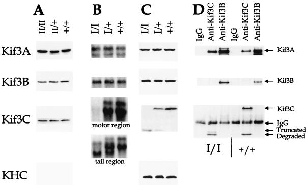
FIG. 2.
Analysis of Kif3CtypeI and Kif3CtypeII mice. (A) Western analysis of Kif3CtypeII mice. Brain lysates from Kif3CtypeII (−/−), Kif3CtypeII (+/−), and wild-type (+/+) littermates were analyzed by sodium dodecyl sulfate-polyacrylamide gel electrophoresis (SDS-PAGE). The duplicate blots were probed with antibodies against Kif3A, Kif3B, and Kif3C. (B) Northern analysis of Kif3CtypeI mice. Total RNA was isolated from the Kif3CtypeI mouse brains and analyzed in a formaldehyde-agarose gel. The duplicate blots were probed with cDNA of Kif3A, Kif3B, and Kif3C. Two different probes, encoding either the motor region or the tail region as indicated, were used for Kif3C. (C) Western analysis of Kif3CtypeI mice. Brain lysates from Kif3CtypeI mice were analyzed by SDS-PAGE. The duplicate blots were probed with antibodies against Kif3A, Kif3B, and Kif3C and a monoclonal antibody, SUK4, for kinesin heavy chain. The antibodies against Kif3C detected a truncated Kif3C protein. (D) Immunoprecipitation-Western analysis of Kif3CtypeI mice. Brain lysates from Kif3CtypeI mice were immunoprecipitated with anti-Kif3C or anti-Kif3B antibodies or immunoglobulin G (IgG) as a control. The immunoprecipitated samples were analyzed by SDS-PAGE. The same blot was probed and reprobed with antibodies against Kif3A, Kif3B, and Kif3C. The samples in the left three lanes were from Kif3CtypeI (−/−) mice and the samples in the right three lanes were from wild-type littermates. (The truncated Kif3C protein in wild-type mice was probably generated from protein degradation.)

