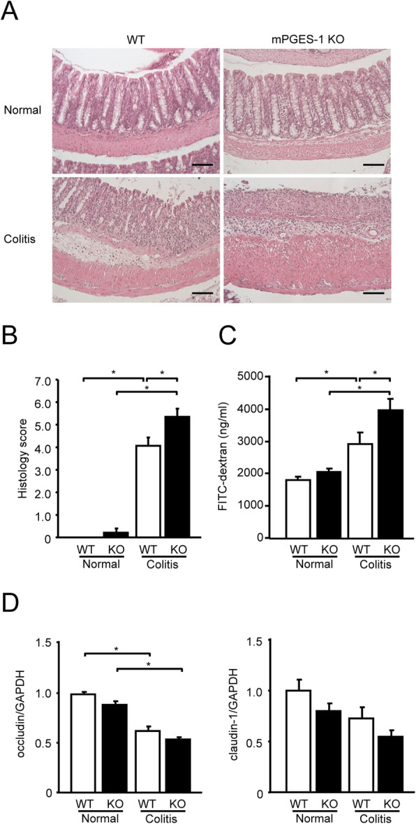
Fig. 2.

Histological analysis of DSS-induced colitis in mPGES-1−/− mice. A Colons of mPGES-1 WT and mPGES-1−/− mice were collected on day 7 after the start of exposure to 1% DSS, and sections were stained with H&E. Results are representative examples adapted with the Swiss roll technique in WT and mPGES-1−/− mice (n = 5 to 17). Scale bar, 100 μm. B Histological scores in WT and mPGES-1−/− mice (n = 5 to 17). Colon sections were examined by a blinded researcher, who calculated the epithelial damage score and inflammatory infiltration score and summed the 2 scores (maximum score: 8). C Intestinal permeability was assessed with FITC-dextran on day 7 after the start of exposure to 1% DSS (n = 9 to 17). *P < 0.05; 2-way ANOVA followed by Tukey multiple comparison test. D Expression of mRNA for the tight junction molecules occludin and claudin-1 in colon from mice treated or not treated with 1% DSS for 7 days were analyzed by real-time RT-PCR. Levels of mRNA expression are shown as the fold induction relative to the expression in WT mice without DSS administration (assigned the value “1”). *P < 0.05; 2-way ANOVA followed by Tukey multiple comparison test (n = 7 to 10)
