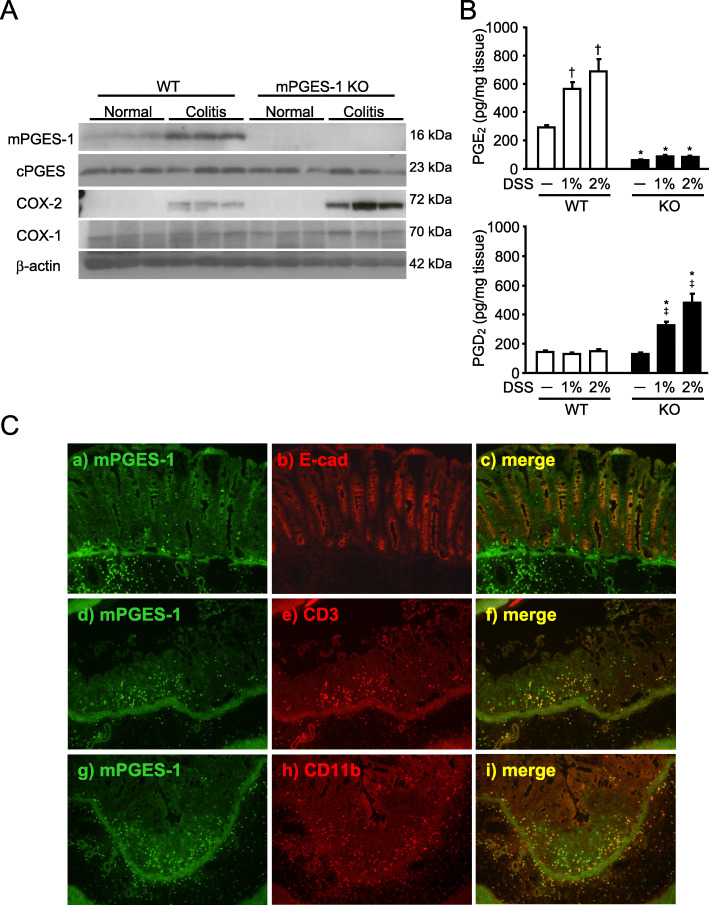
Fig. 5.
mPGES-1 protein expression and prostanoid production in the colon with colitis by DSS. A Expression of protein for PGES and COX isozymes in colon on day 7 after the start of exposure to 1% DSS were analyzed by western blot analysis (n = 3). B The levels of PGE2 and PGD2 in the colon from mice treated or not treated with the indicated dose of DSS for 7 days were measured by ELISA. *P < 0.05 vs WT mice within each day, †P < 0.05 vs non-DSS-treated WT mice, and ‡P < 0.05 vs non-DSS-treated KO mice; 2-way ANOVA followed by Turkey multiple comparison test (n = 3 to 5). C Representative double immunofluorescence staining image of Swiss-roll colon sections of WT mice on day 7 after the start of exposure to 1% DSS. Double staining for mPGES-1 (green) and E-cadherin, CD3 or CD11b (red) showed mPGES-1 immunoreactivity mostly colocalized with an epithelial cell marker, E-cadherin, a T cell marker CD3 and a monocytes/macrophage marker CD11b in the colon

