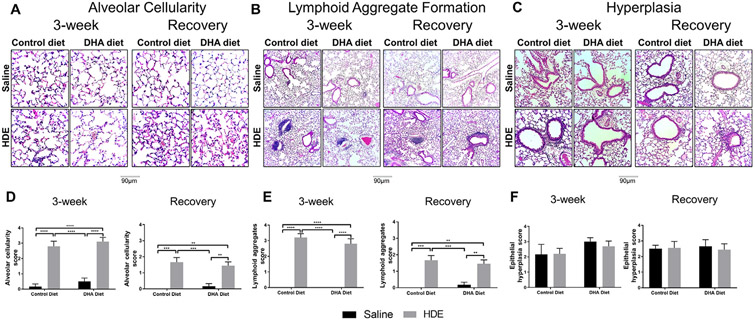
Fig. 4.
Representative images and scoring of lung histopathology in mice receiving DHA diet supplementation and repetitive HDE exposure at three weeks and after the recovery period. Mice were fed DHA or control diets for four weeks before commencing three-week repetitive 12.5% HDE exposure. Lung was fixed with formalin, and paraffin embedded sections were obtained for H & E staining as described in Methods. Lung histopathology was evaluated for alveolar cellularity, lymphoid aggregates, and epithelial hyperplasia both at the end of the three-week HDE exposure (A, B and C, left panels) and recovery period (A, B and C, right panels). H & E-stained lungs were visualized at 200X and 100X, lung pathology scoring was performed based on alveolar cellularity (D), lymphoid aggregates (E) and epithelial hyperplasia (F) as described in Methods. Left and right panels show data for three-week and recovery periods, respectively. Data are mean ± standard error of the mean, n=6 for saline control and DHA alone groups (no HDE exposure) and n=9-10 mice for HDE and DHA+HDE groups. * P<.05, ** P<.01, *** P<.001, **** P<.0001.

