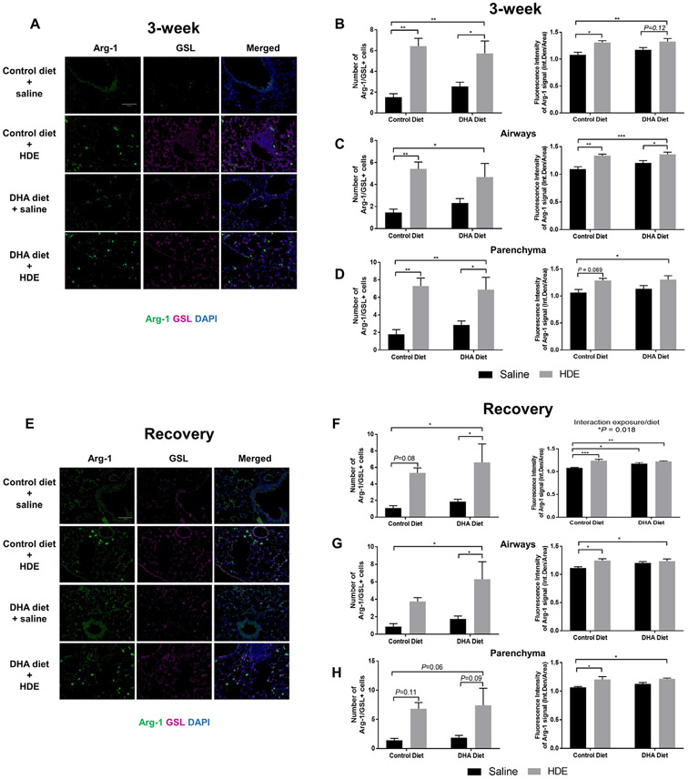
Fig. 8.
Changes in the Arg-1 positive macrophages after repetitive HDE exposure and DHA diet supplementation in the lung at three weeks (A-D) and after the recovery period (E-H). Formalin fixed and paraffin embedded lung sections were stained for Arg-1 and GSL, M2 and M1/M2 macrophage marker in tissues from mice receiving control or DHA diet for four weeks prior to three-week HDE exposure. Images were obtained by epifluorescence microscope Echo Revolve with 20X objective in FITC (Arg-1) and Cy5 (GSL) channels. Between 5-7 images for airways and lung parenchyma were obtained. The fluorescence intensity of the whole image was determined by quantifying integrated density/area in Image J. Quantification of Arg-1 immunofluorescence signal around the airways and parenchyma at three-weeks (C, D) and at the recovery period (G, H) are shown as number of Arg-1/ GSL positive cells and fluorescence intensity. Data are mean ± standard error of the mean. * P<.05, ** P<.01, *** P<.001.

