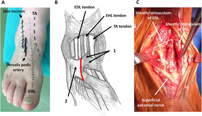
Figure 1.
Preoperative, intraoperative photographs of the ankle. (A) Preoperative design of the skin incision. The skin incision is made 1.5 cm laterally from the EHL. (B) Illustration of retinaculum and tendons around ankle. 1: Superior and inferior subdivisions of the superomedial band of the inferior extensor retinaculum; 2: stem of inferior extensor retinaculum; red line: opening line of the retinaculum. (C) Intraoperative findings of the ankle after opening the retinaculum. The EHL and EDL tendons are covered by sheath and retinaculum. The TA tendon does not appear. Each side of the sheath and retinaculum is marked by threads.

