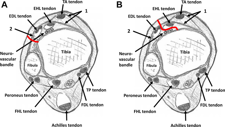
Figure 2.
Axial illustration of the ankle. 1: Superior and inferior subdivisions of the superomedial band of the inferior extensor retinaculum; 2: stem of inferior extensor retinaculum. (A) Red line: approach route to tibia in original anterolateral (Böhler’s) approach; (B) red line: approach route to tibia in modified anterolateral approach. (FDL, flexor digitorum longus; FHL, flexor hallucis longus; TP, tibialis posterior.)

