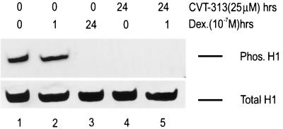
FIG. 2.
Prolonged treatment with dexamethasone and CVT-313 dephosphorylates histone H1 in vivo. Mouse cells were untreated (lane 1), treated with dexamethasone (10−7 M) for 1 h (lane 2), treated with dexamethasone for 24 h (lane 3), treated with CVT-313 (25 μM) for 24 h (lane 4), or pretreated with CVT-313 (25 μM) for 23 h prior to dexamethasone addition for 1 h (lane 5). Total histones were prepared from the nuclei by H2SO4 extraction as described in Materials and Methods. Histones (30 μg) were separated on a 16% acrylamide acid-urea gel, transferred to a nitrocellulose membrane, and analyzed by Western blot analysis using a polyclonal anti-phosphorylated H1 antibody. Equal loading of protein was confirmed by staining the blot with amido black dye to reveal the total H1 present (lower panel).

