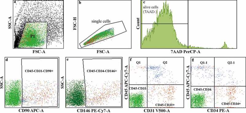
Figure 1.

Flow cytometry analysis of the cellular content of LAT. (a–c) Illustration describing the gating process. The viability assay using 7AAD staining showed 96.6% cell viability in the LAT. The live-cell populations (7AAD−) identified in the LAT used in the in vivo study comprised (d) 1% ASCs (CD45−CD31−CD90+), (e) 1% pericytes (CD34− CD45−CD146+), (f) 3.2% EPCs (CD45−CD34+) and (g) 3.6% endothelial cells (CD45−CD31+).
