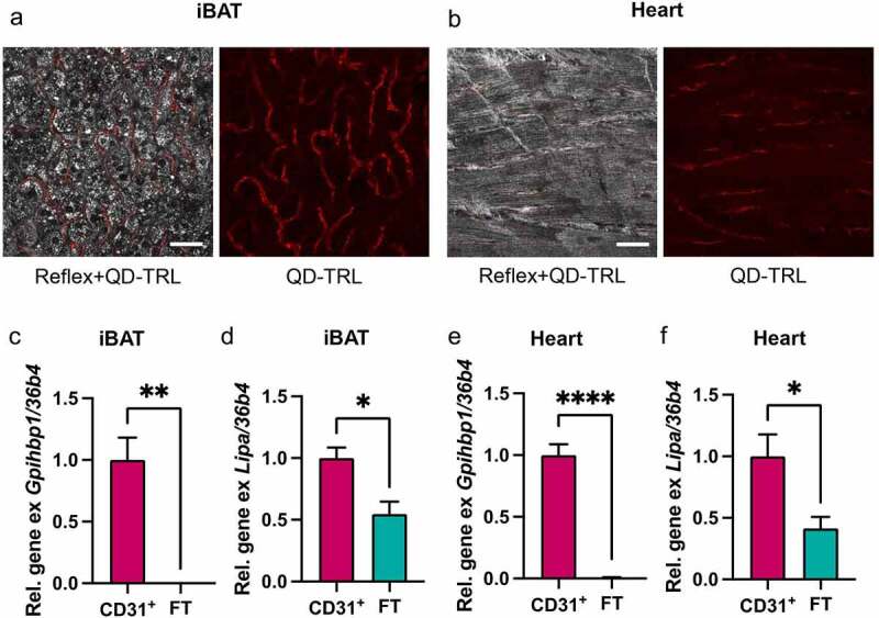
Figure 2.

Endothelial cells of BAT and cardiac muscle take up whole TRL particles, and ECs are equipped to process TRLs in lysosomes
C57BL6/J wildtype mice were injected with recombinant human TRL particles labelled with fluorescent Quantum Dot nanoparticles as described previously [11,22]. 20 min after intravenous injection, organs (BAT, cold activated or heart, standard housing temperature), tissues were dissected imaged as described previously [22,40].(a, b) Confocal microscopy images of QD-labelled TRL particles internalized by BAT (a) and heart tissue (B). Red = QD-TRL. Scale bar = 50 µm.Endothelial cells were isolated from BAT and heart as described previously [12] with minor alterations. Isolation of BAT EC was performed as described, while digestion of heart tissue was performed by incubation of finely minced tissue in PBS containing 10 mM CaCl2, 0.1% (w/v) collagenase I, 0.25% (w/v) collagenase IV, 2.4 U/mL Dispase and 7.5 μg/mL DNAse I for 45 min at 37°C with vigorous shaking and pipetting every 5 min. RNA extraction and qPCR was performed as described previously [22].(c, e) Gene expression of GPIHBP1 and (d, f) Lipa in CD31+ endothelial cells of BAT (c, d) or heart (e, f) isolated by MACS® (n = 4).
