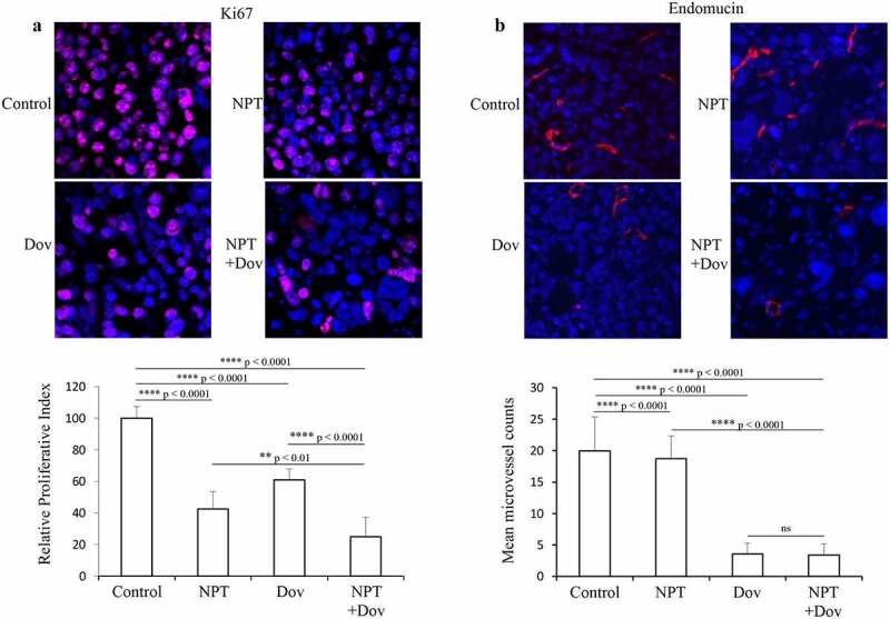
Figure 3.

Dovitinib and nab-paclitaxel reduce tumor cell proliferation and tumor vasculature in GAC cell-derived xenograft: Tumor sections derived from MKN-45 xenografts after 2-week therapy with control, dovitinib, nab-paclitaxel, and nab-paclitaxel plus dovitinib were analyzed by IHC. (a) Tumor cell proliferation was determined after incubating tumor sections with anti-Ki67 antibody followed by counting the Ki67-positive cells from 5 high-power fields (HPF). Cell nuclei stained with Ki67 (red) and DAPI (blue) are illustrated at 20X magnification. ** p < .01; **** p < .0001 by t-test. (b) Microvessel density was determined by incubating with endomucin antibody followed by calculating endomucin positive vessels in 5 HPF. Endomucin positive microvessel (red) and cell nuclei (DAPI, blue) are illustrated at 20X magnification. **** p < .0001 by t-test. The results are displayed as mean values ± standard deviation for each treatment group
