Figure 5.

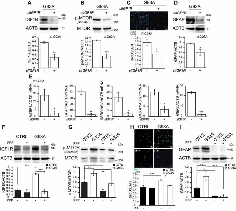
Genetic and pharmacological manipulation of IGF1R in SOD1G93A iPSA reverses proliferation and astrocytic reactivity. G93A iPSA were transfected with siRNA for IGF1R (+) or for Renilla luciferase (-) for 48 h prior to the assays. (A) Representative western blot and quantification of IGF1R protein levels normalized to ACTB (n = 3), (B) p-MTOR (Ser2448) protein level normalized to total MTOR (n = 3). (C) Representative images of CTRL and G93A iPSA stained with BrdU (green) and DAPI (blue), and quantification of cell proliferation (n = 6). (D) Representative western blot and quantification of GFAP protein levels normalized to ACTB (n = 3). (E) HSBP1, GFAP, SERPING1, GGTA1, GBP2 mRNA levels normalized to ACTB (n = 3). CTRL and G93A iPSA were treated with 15 μM PPP (+) or vehicle (-) for 24 h. (F) Representative western blot and quantification of IGF1R protein levels normalized to ACTB (n = 3), (G) p-MTOR (Ser2448) protein level normalized to total MTOR (n = 4). (H) Representative images of CTRL and G93A iPSA stained with BrdU (green) and DAPI (blue), and quantification of cell proliferation (n = 9). (I) Representative western blot and quantification of GFAP protein levels normalized to ACTB (n = 4). P values <0.05 by unpaired Student’s t test (A-E) or by one-way ANOVA test with Sidak’s correction between indicated groups (F-I) are shown
