
Figure 2.
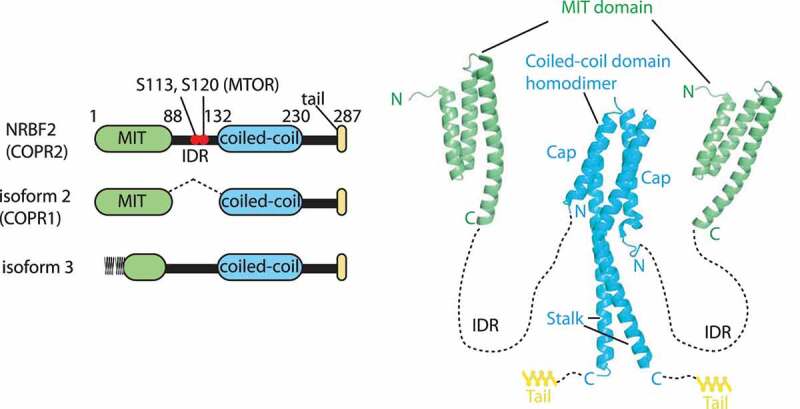
Structures of NRBF2/Atg38. left: schematic representations of NRBF2 and its isoforms (in human residue numbers). The S113 and S120 residues in the intrinsically disordered region (IDR) are phosphorylated by MTOR. Right: structures of the NRBF2 MIT domain (PDB: 4ZEY) and Atg38 coiled-coil domain (PDB: 5KC1). NRBF2/Atg38 makes a stable homodimer via the two segments, the cap and stalk in the coiled-coil domain