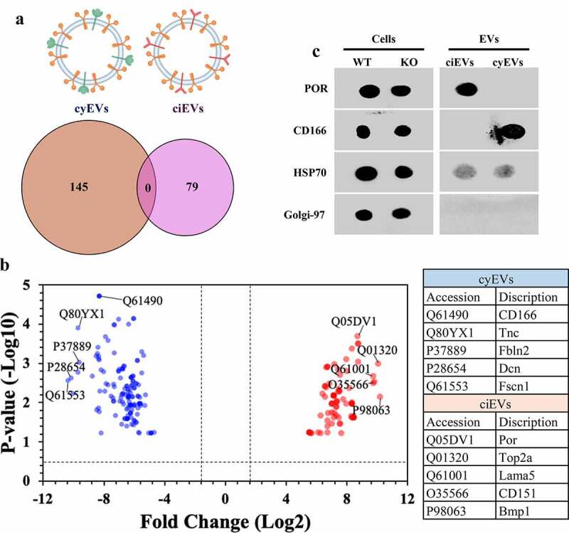
Figure 1.

ciEVs and cyEVs have unique biomarkers
(a) EV isolation from ciliated (wild-type; ciEVs) and non-ciliated (IFT88; cyEVs) cells reveals unique biomarkers. (b) The volcano plot shows the top five distinctive identified biomarkers based on their pvalue and fold-change for cyEVs (blue color) and ciEVs (red color). The dot blot analyses show the expression of the top cyEV and ciEV biomarkers (CD166 and POR, respectively) in isolated EVs. HSP70 and Golgi-97 were used as positive and negative controls for extracellular vesicles, respectively.
