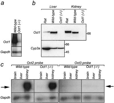
FIG. 2.
Oct1 mRNA and protein analysis. (A) Expression of Oct1 RNA in livers of wild-type and Oct1−/− mice. RPA was performed with 10 μg of total RNA per sample. Oct1- and Gapdh-protected RNA fragments originate from the same gel, and their positions are indicated. Note that the specific activity of the Gapdh probe was 100-fold lower than that of the Oct1 probe. (B) Immunodetection of Oct1 in livers and kidneys of wild-type and Oct1−/− mice. A polyclonal antibody raised against rat Oct1 (which cross-reacts with mouse Oct1) was used on crude membrane fractions of liver (20 μg per lane) and kidney (10 μg per lane). The same blot was incubated with a monoclonal antibody raised against rat cytochrome P450 3a (Cyp3a; expressed only in the liver), used as a protein loading control. Molecular weight marker bands are indicated in kilodaltons. (C) RPA of Oct2 and Oct3 RNA in brains, livers, and kidneys of wild-type and Oct1−/− mice. Experimental details are as described for panel A. Arrows indicate protected fragments of Oct2 (left) and Oct3 (right).

