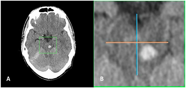
Fig. 5.
Deliniation of anterior versus posterior brainstem injuries. a Axial head computed tomography (CT) slice demonstrating left midbrain traumatic brainstem injury. b Magnified view of area within green box demonstrated in panel a showing midline anterior–posterior line (blue) with perpendicular bisecting line (orange) situating the injury in the posterior half of the brainstem

