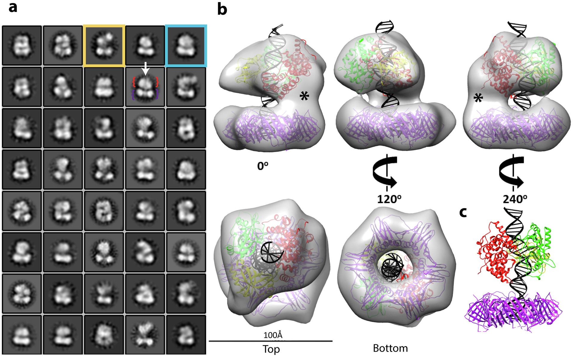
Fig. 7. Structure of DNA-bound PCNA-LigI complex.

Single particle EM analysis of complexes formed by LigI and PCNA were co-purified with non-ligatable nicked DNA. a. 2D classification reveals DNA (white arrow) traversing two separate densities: PCNA (purple bracket) and LigI (red bracket). The boxsize is 210 Å in length and width, the circular mask is 150 Å. Classes show open (yellow box) and stacked rings (blue box) conformations. b. The stacked rings 3D map (~20 Å resolution) of the complex is shown at the indicated rotations. Crystal structures of PCNA (PDB 1AXC) and LigI-DNA (PDB 1X9N) were docked into the lower and upper rings, respectively, with extended nicked DNA. LigI was positioned with its DBD (red) adjacent to the density linking to PCNA. The empty density (*) is attributable to the unstructured N-terminal region of LigI. The LigI AdD (green) mediates a second point of contact with PCNA and the OBD (yellow) completely encircles the nicked DNA. c. A 240° view of the modeled crystal structures.
