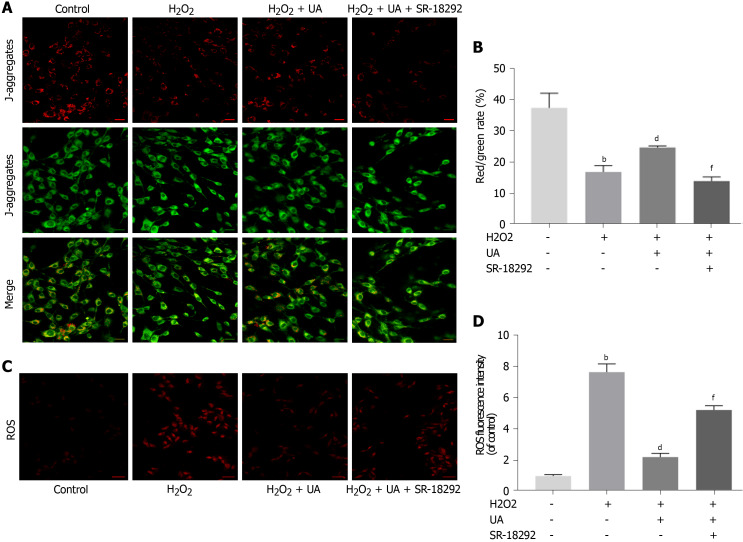
Figure 5.
Mitochondrial membrane potential assay and reactive oxygen species assay. A: Results of mitochondrial membrane potential (MMP) in different groups detected by fluorescence. Red fluorescence represents the mitochondrial aggregate JC-1 and green fluorescence indicates the monomeric JC-1 (scale bar = 50 μm); B: Quantitative analysis of MMP results; C: Results of reactive oxygen species (ROS) in different groups detected by fluorescence. Red fluorescence represents high level of ROS (scale bar = 50 μm); D: Quantitative analysis of ROS results. All data are expressed as the mean ± SD. aP < 0.05, bP < 0.01 compared with control group; cP < 0.05, dP < 0.01 compared with H2O2 group; eP < 0.05, fP < 0.01 compared with H2O2 + UA group. MMP: Mitochondrial membrane potential; ROS: Reactive oxygen species; UA: Urolithin A.

