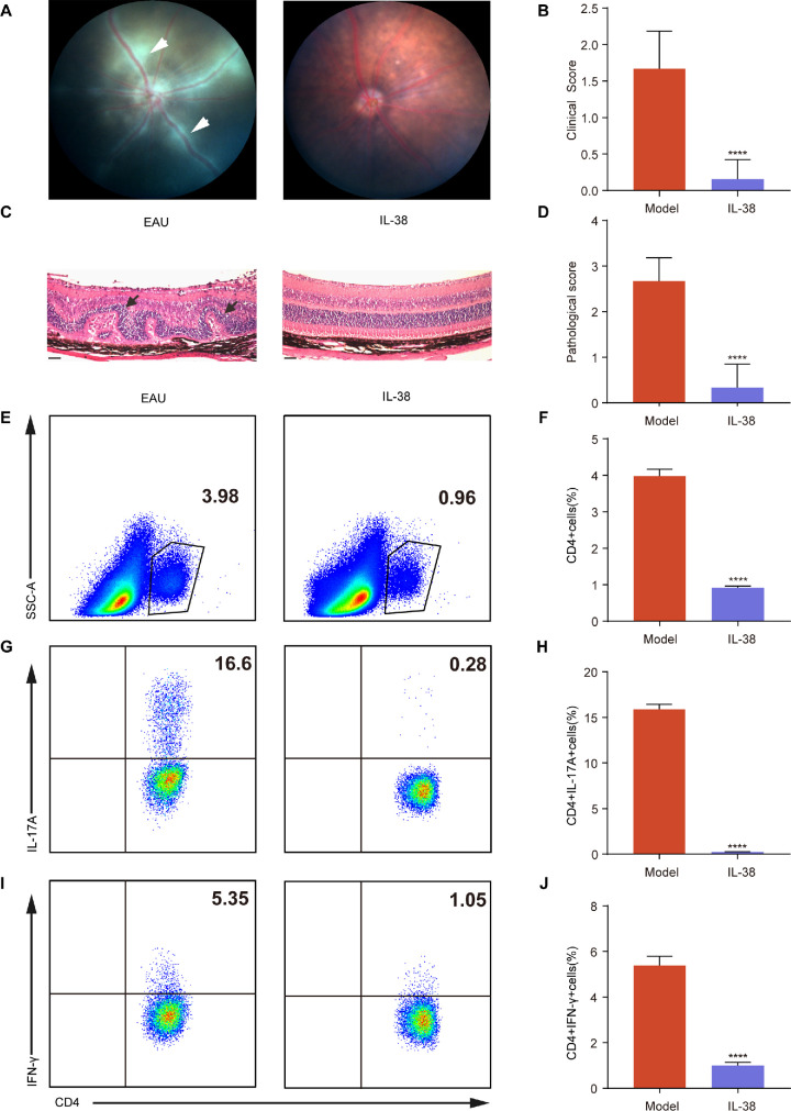
Figure 1.
IL-38-mediated amelioration of EAU and Inhibition of Teff Cell infiltration into the Retina. (A) The representative fundus images of eyes in EAU mice and IL-38-treated mice after immunization at day 14 (n = 6). The white arrows indicate inflammatory exudation and vascular deformation. (B) Clinical scores of EAU mice and IL-38-treated mice (n = 6). (C) The H&E staining of EAU mice and IL-38-treated mice after immunization at day 14 (n = 6). Scale bars, 20 mm. The black arrows indicate retinal folding. (D) Histological scores of EAU model mice and IL-38-treated mice (n = 6). (E–J) The proportion of CD4+ (gated on living cells) E to F, CD4+IL-17A+(Th17) cells (gated on CD4+ cells) G and H, and CD4+IFN-γ+ (Th1) cells (gated on CD4+ cells) (I–J) infiltrated into retina after immunization at day 14 in EAU mice and IL-38-treated mice (n = 6). Data shown as mean ± SD from three independent experiments. Data were analyzed using unpaired two-tailed Student t-tests, ****P < 0.0001.

