Fig. 3.
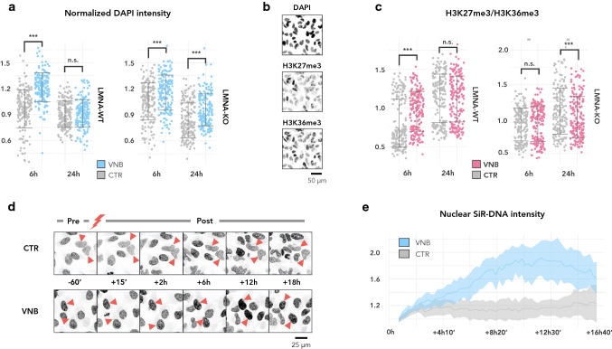
VNB photoporation causes a transient chromatin compaction, which is prolonged in LMNA-KO cells. a Normalized nuclear DAPI intensity plotted per timepoint for LMNA-WT (left) and LMNA-KO cells (right) (***P < 0.001); b Representative example of a staining with DAPI, anti-H3K27me3, and anti-H3K36me3 on CTR cells at 6 h. c Ratio of heterochromatin (H3K27me3) to euchromatin (H3K36me3) plotted per timepoint for LMNA-WT (left) and LMNA-KO (right) (***P < 0.001); d Montage of SiR-DNA stained LMNA-WT cells before photoporation treatment (− 60’) and at several timepoints after photoporation treatment (+ 15’, + 2 h, + 6 h, + 12 h and + 18 h). Control cells were irradiated in the absence of AuNPs; e Line graphs of the mean SiR-DNA signal (± standard error) in VNB-treated cells (VNB) and control cells that were irradiated in the absence of AuNPs (CTR). Per treatment, SiR-DNA signal was normalised to the mean intensity of the first timepoint (+ 15’) post laser irradiation (n = 4 cells per treatment)

