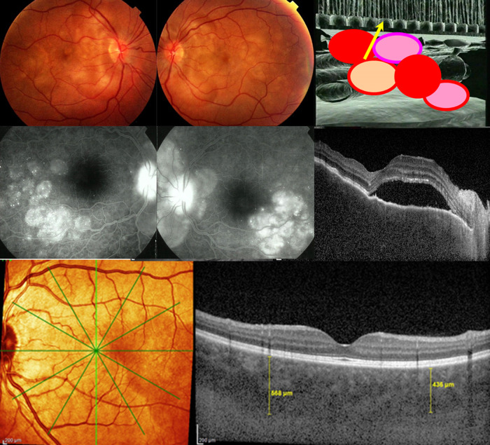
Fig. 2. Signs of initial-onset, acute exudative VKH disease.
a Fundoscopy images show bilateral retinal exudative detachments that resulted from (b, cartoon) choroidal inflammation spill-over into the retina and optical disc. c FA and d OCT images show exudative retinal detachments. e EDI-OCT images show choroidal thickening in excess of 400 µm (yellow vertical lines), in early disease.

