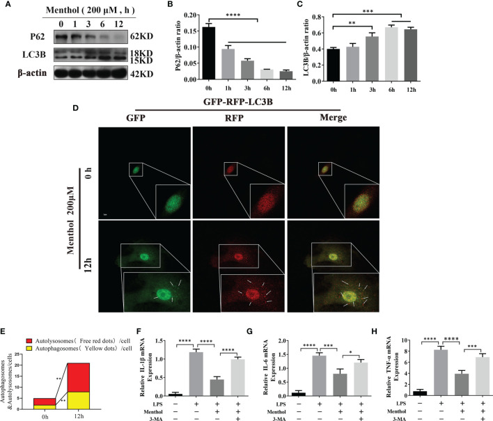
Figure 3.
Menthol alleviates the LPS-induced inflammatory response by activating autophagy in BMECs. The BMECs were treated with 200 μM menthol at different time points (0, 1, 3, 6, and 12h). (A) Western blotting assay for P62, HO-1, and LC3B protein expression levels (n≥3). (B, C) Bar graphs indicate the quantitative results of the corresponding protein bands. β-actin was used as an internal reference to homogenize the protein results. Plasmids carrying mRFP-GFP-LC3B were transfected into BMECs, stably expressed cell were constructed. BMECs treated with 200 μM menthol for different times (0,12h).And images were obtained by confocal microscopy. Yellow spots represent autophagosomes and red spots represent autolysosomes. (D) Results of laser confocal detection of expression changes in BMECs of LC3B constructs. (E) Results of the quantify of autophagosome and autolysosomes formation, data were obtained from 3 independent experiments, each with a quantitative score of 10 cells. (F–H) Results of qRT-PCR for mRNA levels of IL-1β, IL-6, and TNF-α. β-actin was used as an internal reference for homogenization(n≥3). The values are presented as the means ± SEM (*p < 0.05, **p < 0.01, ***p < 0.001 and ****p < 0.0001).

