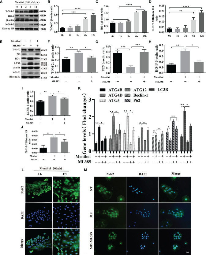
Figure 7.
Menthol can promote the expression of autophagy-related genes by activating the Nrf-2/HO-1 signaling axis. The BMECs were treated with 200 μM menthol at different time points (0, 1, 3, 6, and 12h). (A) Western blotting for the protein expression levels of T-Nrf-2, HO-1, N-Nrf-2 and Histone H3 (n≥3); T-Nrf-2 represents total intracellular Nrf-2 and N-Nrf-2 represents Nrf-2 in the nucleus. (B–D) Bar graphs indicate the quantitative results of the corresponding protein bands. Then 200 μM menthol and ML385 (inhibitor of Nrf-2, 5 μg/ml) were pretreated with bovine mammary epithelial cells for 1 h. LPS at 5 μg/ml was added for 12 h. (E) Protein expression levels of T-Nrf-2, p62, HO-1, LC3B, N-Nrf-2 and Histone H3 were detected by western blotting (n≥3); T-Nrf-2 represents total intracellular Nrf-2, and N-Nrf-2 represents Nrf-2 in the nucleus. (F–J) The bar graph indicates the quantitative results of the corresponding protein bands. β-actin was used as an internal reference protein to homogenize the protein results. Histone H3 was used as the internal reference protein in the nucleus. (K) Results of qRT-PCR assay for mRNA levels of ATG4B, ATG4D, ATG5, ATG12, Beclin-1, P62, LC3B (n≥3). The mRNA expression of β-actin was used to normalize the mRNA expression of the above proteins. The BMECs were treated with 200 μM menthol at different time points (0, and 12 h). (L) Immunofluorescence staining to detect the entry of Nrf-2 into the nucleus, nucleus (blue), and Nrf-2 (green). Then 200 μM menthol and ML385 (inhibitor of Nrf-2, 5 μg/ml) were pretreated with bovine mammary epithelial cells for 1 h LPS at 5 μg/ml was added for 12 h. (M) Immunofluorescence staining to detect the entry of Nrf-2 into the nucleus, nucleus (blue), and Nrf-2 (green). The values are presented as the means ± SEM (ns means no difference, *p < 0.05, **p < 0.01, ***p < 0.001, and ****p < 0.0001).

