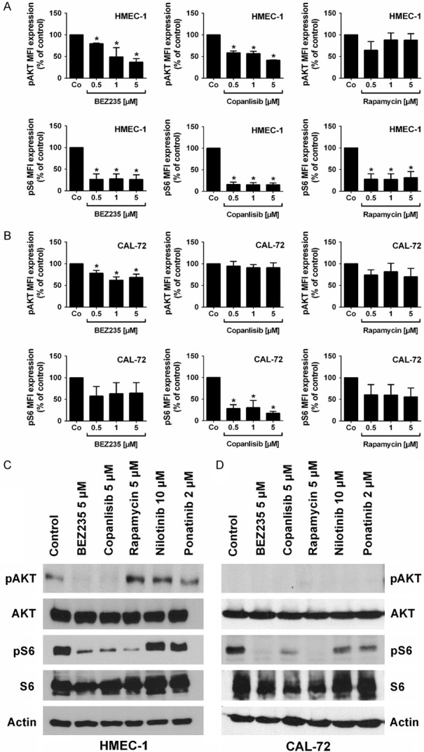
Figure 5.

Effects of BEZ235, copanlisib and rapamycin on expression of phosphorylated (p) AKT and S6 in endothelial and osteoblastic niche cells. (A, B) Flow cytometric evaluation of expression of pAKT and pS6: HMEC-1 cells (A) and CAL-72 cells (B) were incubated in control medium (Co) or in medium containing various concentrations of BEZ235 (0.5-5 μM), copanlisib (0.5-5 μM) or rapamycin (0.5-5 μM) at 37°C for 4 hours. Then, cells were permeabilized and stained with antibodies against pAKT (S473) and pS6 (S235/236). Expression of pAKT and pS6 was determined by flow cytometry. Results show median fluorescence intensity (MFI) values expressed as percent of control and represent the mean ± SD from 3 independent experiments. Asterisk (*): P<0.05 compared to control. (C, D) Effects of BEZ235, copanlisib and rapamycin on expression of phosphorylated (p) AKT and S6 in endothelial and osteoblastic niche cells as determined by Western blotting. HMEC-1 cells (C) and CAL-72 cells (D) were incubated in control medium or in medium containing BEZ235 (5 μM), rapamycin (5 μM), copanlisib (5 μM), nilotinib (10 μM), or ponatinib (2 μM) at 37°C for 4 hours. Then, cells were subjected to Western blot analysis as described in text using antibodies directed against pAKT, total AKT, pS6, total S6 or actin. The figure show data from one typical Western blot experiments. Corresponding results were obtained in two other Western blot experiments.
