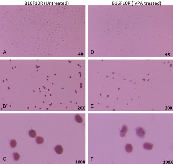
Figure 11.

Morphological changes in drug-resistant melanoma (B16F10R) cells upon VPA treatment. B16F10R cells were treated with VPA (2 mM) for 24 h. Image of untreated B16F10R in objectives magnification of, (A) 4×; (B) 20×; and (C) 100×; after 24 h after VPA treatment: (D) 4×; (E) 20×; and (F) 100×. Cells were stained using Pap stain method. Cell morphology was captured under a light microscope (Olympus CX41).
