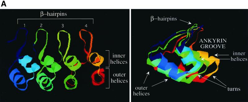
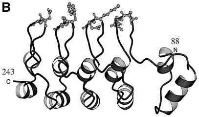
FIG. 2.
Secondary-structure prediction of the ankyrin repeat domain of RFXANK. (A) Schematic representation of the secondary-structure elements of the ankyrin repeats in a three-dimensional view. Four ankyrin repeats represent only a general ankyrin domain structure. In the left scheme, β-hairpin loops form the loop structures above the two planes of helices (inner and outer helices). The right scheme represents the same three-dimensional structure from a different perspective (as viewed from the first ankyrin repeat towards the last). The L-shaped structure appears, forming the ankyrin groove. β-Hairpin loops, turns, and inner and outer helices form four different surfaces of the ankyrin repeat domain and are depicted with arrows. (B) Model structure of RFXANK residues 88 to 243. The ankyrin repeat domain of RFXANK is depicted as a ribbon structure, with two exposed variable residues at the very tip of each of the four β-hairpin loops highlighted. This figure was generated with Molscript (19). N, amino terminus; C, carboxy terminus.


