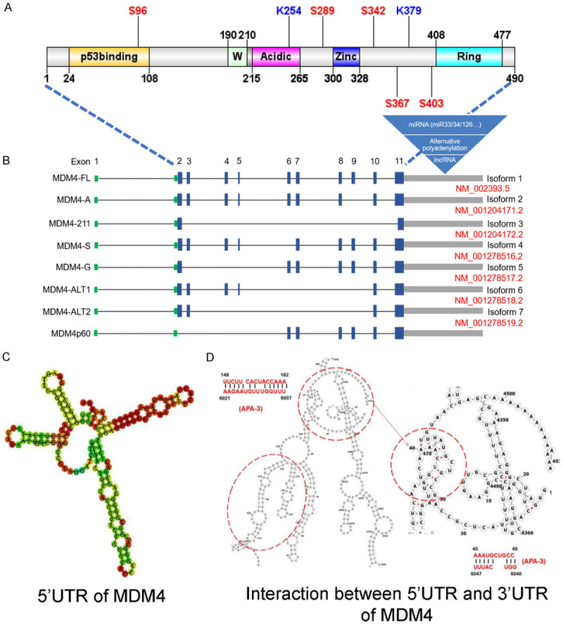
Figure 1.

Molecular structures of MDM4 gene and protein. A. A diagram of MDM4 includes known domains and the variants present in this manuscript. B. The 5’UTRs are shown as green bars and the 3’UTRs are shown as grey bars. Separated by introns shown by black lines, exons are indicated by blue solid rectangles. Seven transcripts were listed in NCBI database with indicated accession numbers and one currently identified variant has no accession number. Multiple miRNAs, APA, UPF1 and STAU1 binding motifs were found in 3’UTR region of MDM4 mRNA. C. RNA secondary structure prediction for 5’UTR of MDM4 using online RNA fold program. D. Detailed information about 5’ and 3’UTR interacting regions predicted in MDM4 mRNA.
