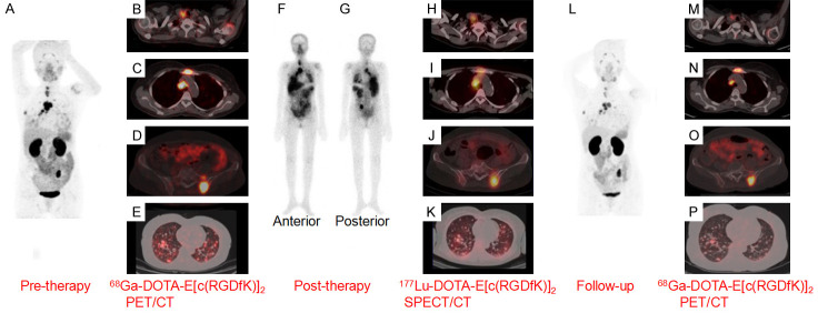
Figure 12.
A 54-year-old woman with papillary thyroid carcinoma who developed TENIS syndrome after receiving 500 GBq of 131I in cumulative doses, was administered with 177Lu-DOTA-E[(cRGDfK)2] therapy. 68Ga-DOTA-E[(cRGDfK)2] PET/CT was performed to evaluate disease extent and for pre-therapy assessment. (A) Pre-therapy, the maximum intensity projection (MIP) image with 68Ga-DOTA-E[(cRGDfK)2] PET scan, transaxial fused PET/CT images showed increased tracer uptake in the (B) thyroid remnant, (C) cervical lymph nodes (D) mediastinal lymph node, lytic skeletal lesions with soft tissue component in the sternum and left iliac bone, (E) multiple lung nodules. Post-therapy WBS in (F) anterior and (G) posterior views revealing the overall distribution of 177Lu-DOTA-E[(cRGDfK)2] and transaxial fused SPECT/CT images (H-K) showing tracer uptake at sites corresponding to 68Ga-DOTA- E[(cRGDfK)2] -avid lesions. (L) Post-therapy follow-up 68Ga-DOTA- E[(cRGDfK)2] PET/CT MIP image and transaxial fused PET/CT images showed tracer uptake in the (M) thyroid remnant with cervical lymph nodes, (N) mediastinal lymph node, lytic skeletal lesions with significant reduction in soft tissue component in the sternum, (O) left iliac bone and (P) multiple lung nodules, suggesting response to therapy. Adapted from Ref [161].

