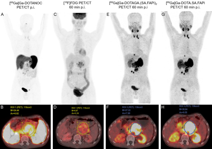
Figure 10.
36-year-old male diagnosed with grade III pancreatic neuroendocrine tumor, the extent off cancer involved primary tumor in the pancreas and left supra clavicular lymph node. MIP and PET/CT fused axial images 1 h p.i. of (A, B) [68Ga]Ga-DOTANOC; (C, D) [18F]FDG; (E, F) [68Ga]Ga-DOTAGA.(SA.FAPi)2 and (G, H) [68Ga]Ga-DOTA.SA.FAPi. Highest primary tumor uptake in pancreas: (H) [68Ga]Ga-DOTA.SA.FAPi PET/CT (SULpeak: 30.84); (F) [68Ga]Ga-DOTAGA.-(SA.FAPi)2 (SULpeak: 27.22). Left supraclavicular visible on (E) [68Ga]Ga-DOTAGA.(SA.FAPi)2 and (G) [68Ga]Ga-DOTA.SA.FAPi.

