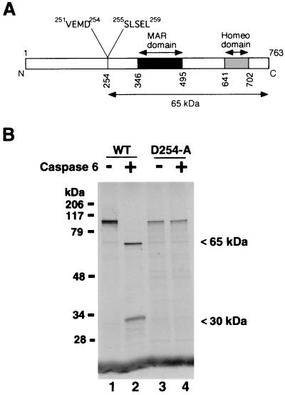
FIG. 5.
Caspase 6 cleaves after aspartate 254 in SATB1. (A) Schematic representation of various known functional domains in SATB1. Black and gray boxes, MAR-binding domain and homeodomain (13), respectively. The caspase 6 cleavage site is located at amino acid (aa) position 254. The caspase 6 recognition sequence (aa 251 to 254) and the N-terminal sequence of the 65-kDa cleavage product (aa 255 to 259) are indicated. (B) VEMD-to-VEMA mutation abolishes the cleavage by caspase 6. Wild-type SATB1 (lanes 1 and 2) and the caspase-resistant mutant SATB1-D254A (lanes 3 and 4) were transcribed and translated in vitro and digested with recombinant activated caspase 6 (lanes 2 and 4), as described in Materials and Methods. The intact proteins and fragments were separated by SDS-4 to 15% gradient PAGE (Bio-Rad) and visualized by autoradiography. Positions of the cleavage products are indicated on the right.

