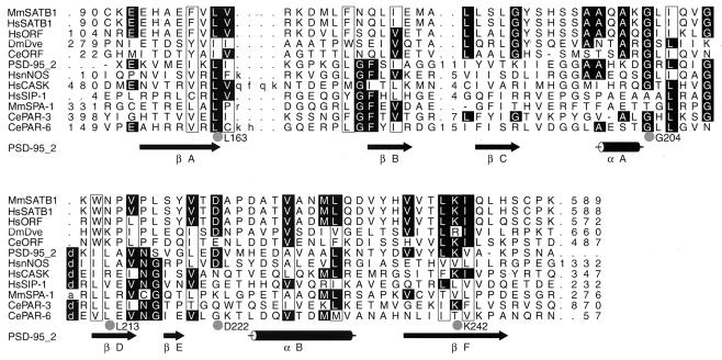
FIG. 9.
SATB1 dimerization domain is homologous to PDZ domains. Shown is an HMM-generated alignment of the dimerization domain of SATB1 and related sequences (MMSatb1 to CeORF) and selected PDZ domains (PSD-95 2 to CePAR-6). Columns containing residues that are conserved in 6 or more of the 12 sequences are highlighted. Dots, columns that are most conserved (10 of 12 sequences) and residues that might be functionally important owing to their spatial proximity on the surface of the PDZ domain based on the alignment in the context of the three-dimensional structure. Hydrophobic columns are boxed. Numbers denote the numbers of amino acids that are not shown. Arrows and cylinders, β-strands and α-helices taken from the nuclear magnetic resonance structure of the second PDZ domain of PSD-95 (PSD-95–2; PDB-RCSB code 1QLC). Amino acids in lowercase within the alignment correspond to residues aligned to the insert state of the HMM. The sequences shown are MmSATB1 (NP 033148), HsSATB1 (NP 002962.1), HsORF (Homo sapiens hypothetical protein KIAA1034; BAA82986.1), DmDve (CAA09729.1), CeORF (hypothetical protein ZK1193.5; T27710), PD-95–2 (second PDZ domain of PSD-95), HsnNOS (P29475), HsCASK (AAB88125), HsSIP-1 (AAB53042), MmSPA-1 (BAA01973), CePAR-3 (T34302), and CePAR-6 (T43216). Species abbreviations are as follows: Mm, Mus musculus; Hs, H. sapiens; Ce, C. elegans; Dm, D. melanogaster.

