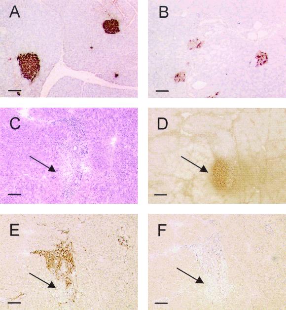
FIG. 5.
Immunohistochemical determination of autoimmune diabetes in APNG−/− mice 8 months after treatment with STZ. (A and B) Formalin-fixed sections from untreated (A) and STZ-treated (B) APNG−/− mice were stained for insulin as described in Materials and Methods. (C to F) CD4+ and CD8+ immunostaining was carried out on frozen sections. Hematoxylin-eosin (C) and insulin (D) staining indicate the position of the islet in the section, while staining for the specific lymphocyte markers shows evidence of CD4+ (E) but not CD8+ (F) lymphocytic invasion in the islet. The more porous nature of the frozen sections compared to the formalin-fixed sections made them unsuitable for quantitative assessment of insulin content by this method. Arrows indicate the position of the islet in the section. Bar, 100 μm.

