Fig. 3.

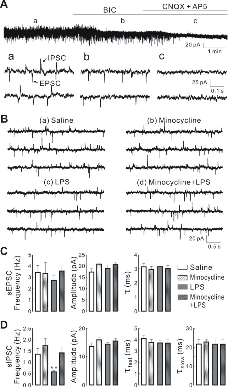
Minocycline inhibits LPS-induced decrease in the frequency of spontaneous inhibitory postsynaptic currents (sIPSCs) in PVN-RVLM neurons. Spontaneous excitatory postsynaptic currents (EPSCs) and IPSCs recorded from PVN-RVLM neurons. (A) top: blockade of upward current by antagonists of GABAA receptor (20 μM Bic; b) and downward current by ionotropic glutamate receptor anta-gonist (50 μM AP5 and 20 μM CNQX; c). Bottom: spontaneous EPSCs (downward) and IPSCs (upward) at expanded time scale before (a), during treatment with BIC (b), and BIC and CNQX + AP5 (c) at a holding potential of −62 mV. (B) Representative traces of spontaneous synaptic currents recorded in the PVN-RVLM neurons derived from saline (a), minocycline (b), LPS (c), and minocycline + LPS groups (d). The synaptic currents were recorded in normal ACSF and at resting membrane potential. All the traces are continuous within each set of records. (C and D) summary bar graphs showing the mean frequency, amplitude, and decay time constants of sIPSCs and sEPSCs in PVN-RVLM neurons obtained from saline (n = 14), minocycline (n = 13), LPS (n = 15), and minocycline + LPS group (n = 15), respectively. Values are mean SEM. **P < 0.01 by one-way ANOVA, followed by the Newman-Keul’s multiple comparison test. τfast and τslow represent fast and slow decay time constants, respectively.
