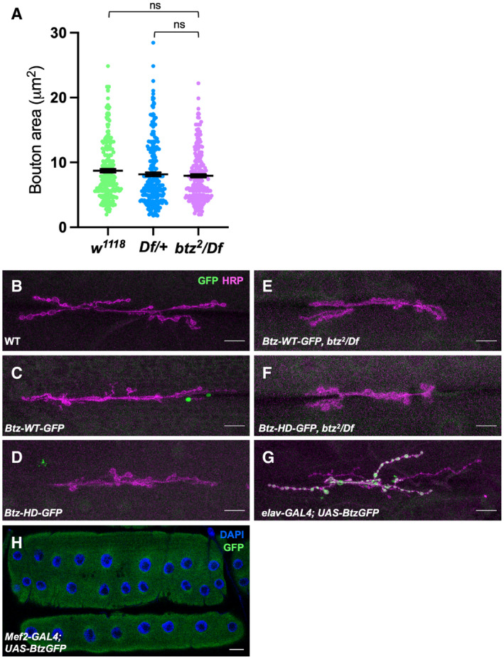
Figure EV1. Btz protein localization at the NMJ.

-
AQuantification of the area of individual synaptic boutons at the NMJ on muscles 6/7 in segment A3 in w1118 and Df(3R)BSC497/+ controls and in btz2 /Df(3R)BSC497 larvae. btz mutants show no significant reduction in bouton area compared to the controls. ns, not significant by Mann–Whitney test. n = 248 boutons from 6 NMJs (w1118 ), 262 boutons from 6 NMJs (Df/+), or 246 boutons from 6 NMJs (btz/Df). Error bars show mean ± SEM.
-
B–HLarval NMJs on muscle 6/7 in segment A3 stained with anti‐HRP (magenta in B‐G), DAPI (blue in H) and anti‐GFP (green). (B) Canton S control; (C) Btz‐WT‐GFP; (D) Btz‐HD‐GFP; (E) Btz‐WT‐GFP; btz2 /Df(3R)BSC497; (F) Btz‐HD‐GFP; btz2 /Df(3R)BSC497; (G) elav‐GAL4; UAS‐Btz‐GFP; (H) Mef2‐GAL4; UAS‐Btz‐GFP. Tagged Btz‐WT and Btz‐HD expressed from the btz promoter fell below our level of detection, even in the absence of endogenous btz. GFP‐tagged Btz overexpressed in neurons with elav‐GAL4 was localized to synaptic boutons, while GFP‐tagged Btz overexpressed in muscle with Mef2‐GAL4 was distributed throughout the muscle. Scale bars, 30 μm.
Source data are available online for this figure.
