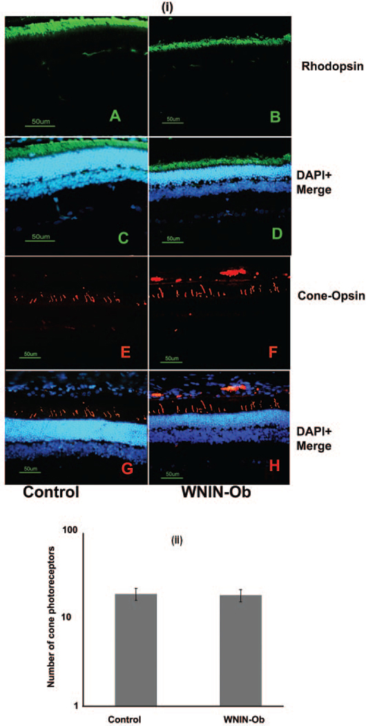
FIGURE 3.
Immunohistochemical evaluation of rod and cone photoreceptor markers in WNIN-Ob rat compared with controls at 12 months. (i) Retinal sections of (A, C, E, G) control and (B, D, F, H) WNIN-Ob rats were labeled with (A–D) rhodopsin and (E–H) cone opsin. Nuclei are labeled with DAPI. Scale bar, 50 μm. (ii) Histogram demonstrates the number of cones in WNIN-Ob and control rat retinas. Values represent mean ± SD of three independent observations (P = 0.2).

