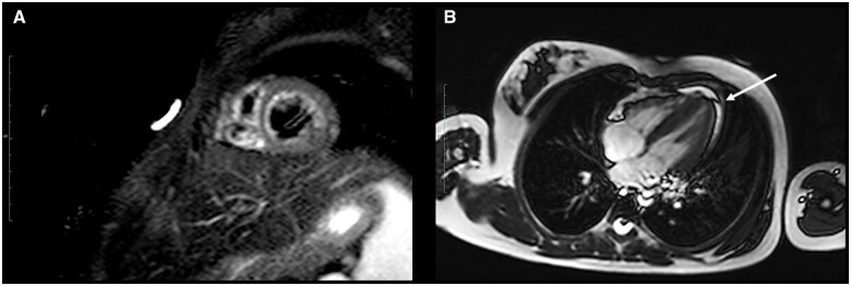
Figure 4.
Cardiac magnetic resonance imaging scans of Patient 1. (A) Short inversion time inversion-recovery sequences showed myocardial signal hyperintensity of the left ventricle, suggesting interstitial oedema. Absence of late gadolinium enhancement suggestive of focal myocardial necrosis/fibrosis. (B) Thin flap of pericardial effusion along the inferior wall of the left ventricle (arrow).

