Figure 3.
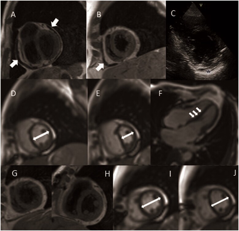
Cardiac magnetic resonance study of Case 3—(A–I) Chronic pericardial constriction, previous to pericardiectomy. Black blood T1 weighted Turbo Spin Echo showing thickening of the pericardium (A, B). (C) Echocardiography, in parasternal short axis view, showing bright and thickened pericardium (blue star). (D–F) Functional assessment of ventricular inter-dependence (short axis in midcavity and long-axis view): (D) normal septal position during expiration, (E) marked leftward septal shift after deep inspiratory effort (white arrows demonstrate the relative change in cavity size due to the septal shift); (F) evidence of septal bounce in long-axis four-chamber views. (G–J) Cardiac magnetic resonance study after pericardiectomy. (G and H) Black blood T1 weighted turbo spin echo confirmed successful partial pericardiectomy. Real-time free breathing: end-expiratory (I) and end-inspiratory (J) septal position, revealing almost completely resolution of constriction signs.

