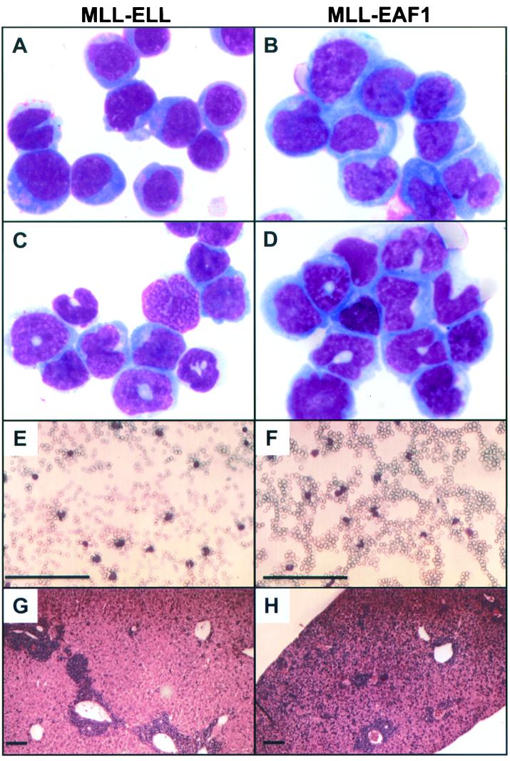
FIG. 5.
Morphology of the neoplastic cells in MLL-ELL (left column) and MLL-EAF1 leukemic mice (right column). Wright-Giemsa staining of BM cytospin preparation (A, B, C, and D). (A) MLL-ELL mouse with acute monoblastic leukemia (poorly differentiated). (B) MLL-EAF1 mouse with acute monocytic leukemia (differentiated). (C and D) MLL-ELL and MLL-EAF1 mice, respectively, with acute myelomonocytic leukemia. (E and F) PB smears showing circulating blast cells. (G and H) Histological analysis of the liver showing infiltration by leukemia cells. Bar, 300 μm.

