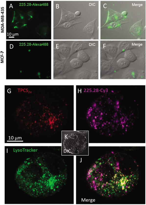
Fig. 2.
Specific binding of mAb 225.28 and co-localization of TPCS2a, mAb 225.28-Cy3 and LysoTracker. (A–F) MDA-MB-435 and MCF-7 cells were incubated with fluorochrome-linked 225.28 for 4 h prior to examination by epi-fluorescence microscopy. (G–K) MDA-MB-435 cells incubated for 18 h in medium containing (G) TPCS2a (1 μg ml−1) and (H) 225.28 mAbs labeled by cyanine 3 (Cy3) (30 nM), washed and chased in fresh medium for 4 h prior to examination by confocal microscopy. (I) LysoTracker Green (1 μM) was added 1 h prior to investigation. (J) Merge of TPCS2a, 225.28-Cy3 and LysoTracker Green. (K) Corresponding DIC micrograph.

