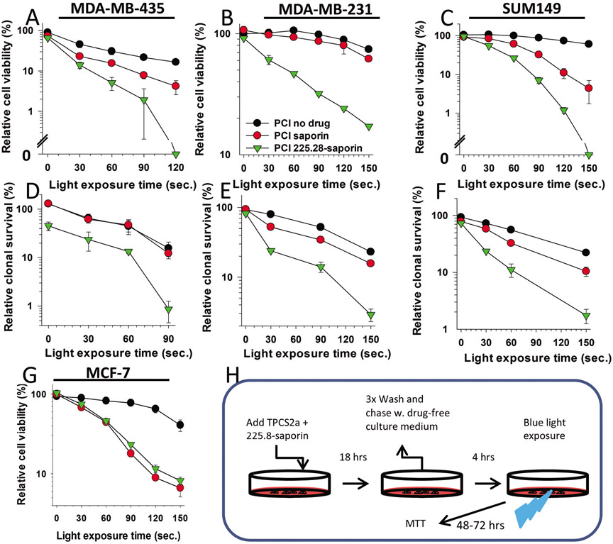
Fig. 3.
Specific and efficient cytotoxic effects induced by PCI of 225.28-saporin in CSPG4-expressing TNBC cell lines. (A), MDA-MB-435, (B) MDA-MB-231 or (C) SUM149 cells were incubated with TPCS2a (PCI no drug), or TPCS2a in combination with streptavidin–saporin or 225.28-saporin (PCI), chased in drug-free medium for 4 h and illuminated as indicated on the X-axis. Relative cell viability was examined using the MTT assay 48 h post illumination. The concentration of TPCS2a was 0.2 μg ml−1 for MDA-MB-231 and MDA-MB-435 and 0.05 μg ml−1 for SUM149. The concentration of toxins was 0.1 nM for MDA-MB-231 and 1 nM for MDA-MB-435 and SUM149. (D–F) Clonogenic survival in (D) MDA-MB-435 (E) MDA-MB-231 or (F) SUM149 cells assessed 10–14 days post light exposure. (G) CSPG-negative MCF-7 control cells after treatments as described in A–C. Each figure shows a single representative experiment out of three independent experiments. Bars = SD of three technical replicates. (H) Experimental protocol of PCI of 225.8-saporin is the same as for PCI of saporin. For PCI no drug (TPCS2a + light only), TPCS2a was incubated alone.

