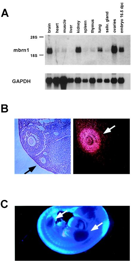
FIG. 2.
Expression of mouse Brainiac 1 in adult mouse tissues and the developing embryo. (A) Northern blot analysis of murine tissues reveals specific expression of a 2-kb transcript. (B) Expression of mouse Brainiac 1 in adult mouse ovaries analyzed by in situ hybridization. The dark-field micrograph shows the expression of mouse Brainiac 1 in follicular granulosa cells in a stage-specific fashion: later-stage follicles with multiple layers of granulosa cells show strong expression of mouse Brainiac 1 (white arrow), while the earlier-stage follicles with single layers of granulosa cells show no expression (black arrow). Control experiments with sense probes were negative (data not shown). (C) Whole-mount in situ hybridization at 12.5 d.p.c. of gestation shows strong expression of mouse Brainiac 1 in the limb buds (arrows).

