Fig. 1.
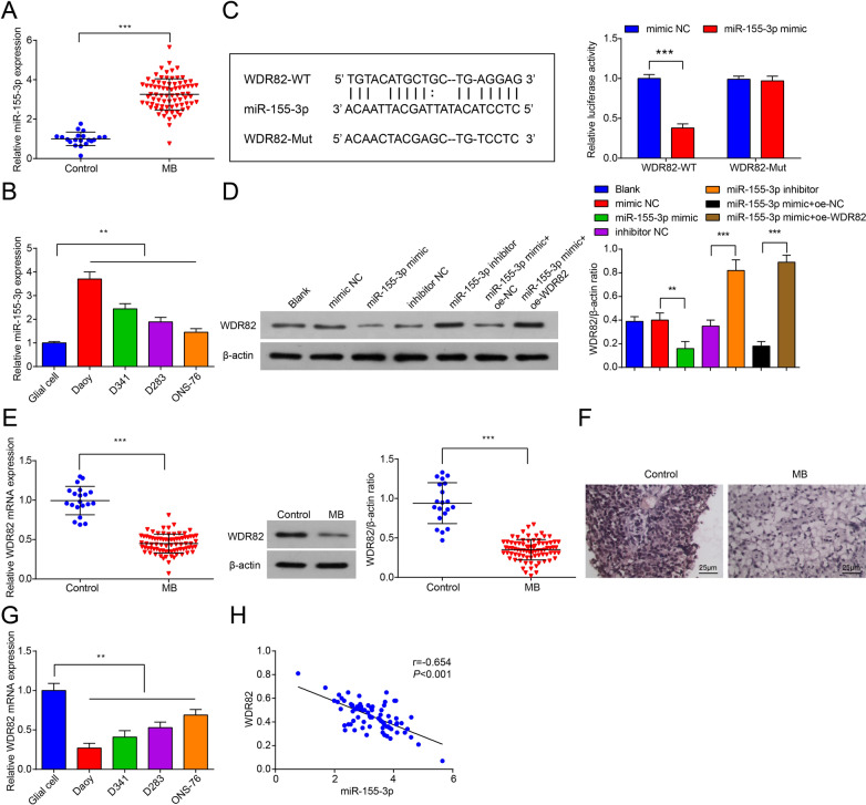
Increase in miR-155-3p level in MB tissues. A MiR-155-3p expression in MB tissues (n = 79) and normal cerebellar tissues (n = 20); B MiR-155-3p expression in Daoy, D341, D283, ONS-76 cells and glial cells; C a dual-luciferase reporter showed significant reduction of luciferase activity of the wild-type, and luciferase activity was restored by the mutant sequence; D WDR82 levels in Daoy cells after transfection; E WDR82 expression in MB tissues (n = 79) and normal cerebellar tissues (n = 20); F WDR82 immunohistochemical staining in MB tissues and normal cerebellar tissues (× 400); G WDR82 expression in Daoy, D341, D283, ONS-76 cells and human glial cells; H the correlation of miR-155-3p and WDR82 mRNA expression in MB tissues. Data were presented as mean ± SD. Statistical analysis was by Student’s t test or one-way ANOVA, Pearson analysis was conducted to determine the correlation between miR-155-3p and WDR82 mRNA expression. **P < 0.01; ***P < 0.001

