Fig. 2.
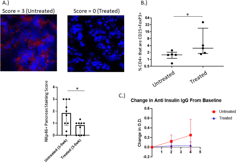
A. Immunohistochemistry of Pancreas. Cryosections of pancreata from NOD mice that were treated with P histicola for 3–5 weeks were stained for the presence of NKp46 and scored for intensity on a scale from 0 to 3. Treatment with P. histicola significantly decreased the intensity for NKp46 staining (p < 0.05). B. Regulatory T cells. NOD mice were treated with P. histicola (Treated) for between 3 and 5 weeks, and pancreatic lymph nodes extracted. % of CD4+ cells that are CD25%FoxP3+ cells significantly increased in the group treated with P. histicola (p < 0.05). Percentages were obtained by first gating on CD4+ cells, then generating dot plots of CD25 vs FoxP3. C. Levels of anti insulin IgG. Circulating levels of anti insulin IgG were evaluated at baseline, 2 weeks of treatment with P. histicola, and 4 weeks of treatement with P. histicola. The change in O. D, from baseline is plotted above and the mice that were treated with P. histicola (n = 10) had much less of an increase in anti insulin IgG than the untreated mice (n = 10)

