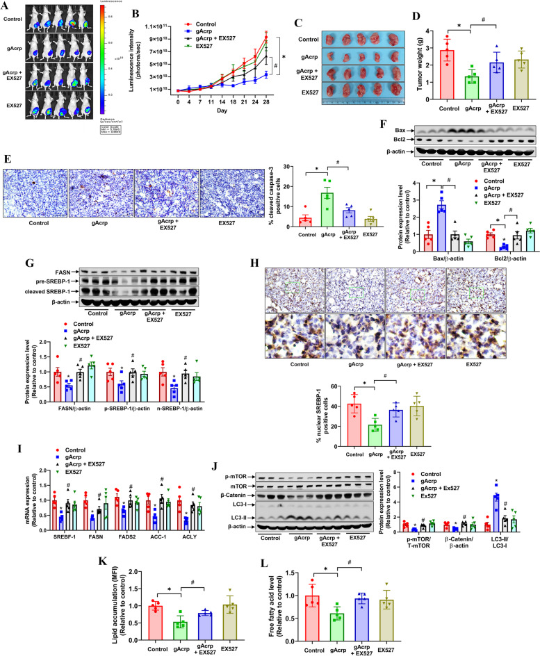
Fig. 7.
Role of SIRT-1 in adiponectin modulation of in vivo breast tumor lipid metabolism and growth. MDA-MB-231-luc orthotopic breast tumors were generated in BALB/c nude mice, followed by treatment with gAcrp alone or gAcrp in combination with EX527 for 28 days. A and B Luminescent images of tumors (A) and tumor growth rate were monitored by luminescent in vivo imaging during treatment (B). C and D Tumor tissues were harvested after 4 weeks of treatment. Isolated tumors were captured at the end of experiment (C) and tumor weight was recorded (D). E Tissue section was prepared, and cleavage of caspase-3 was examined by immunohistochemistry (IHC). The percentage of cleaved caspase-3 positive tumor cells was determined by Image J software. F The expression levels of Bax and Bcl2 were measured by western blot analysis. The representative images from 3 mice each group were shown along with blot quantification for all collected tumor tissues. G The expression levels of FASN and SREBP-1 were analyzed by immunoblotting analysis. H SREBP-1 was detected in tumor tissues by IHC. The proportion of nuclear SREBP-1 positive cells were presented in bar diagram. Scale bar: 100 μm. I The mRNA levels of SREBP-1, FASN, ACC-1, FADS2, and ACLY in tumor tissues were measured by RT-qPCR. J The protein levels of p-mTOR, mTOR, β-catenin, and LC3I/II were determined by western blot analysis. K-L Single cells were isolated from tumor tissues by incubating with collagenase solution. K Tumor cells were incubated with Bodipy 493/503 for 15 min at 37oC, followed by flow cytometry analysis. L The free fatty acid level was measured in tumor cells and normalized to tumor cell number

