. 2021 Oct 6;3(1):44–55. doi: 10.1039/d1cb00135c

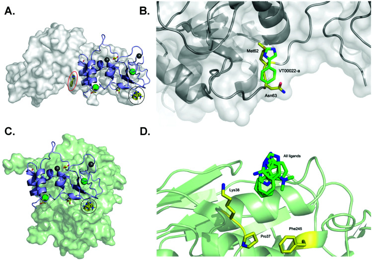
Fig. 3. SARS-CoV-2 nsp10 and all four fragment hits modelled onto either nsp10–nsp14 or nsp10–nsp16 complexes. In all panels, nsp10 is shown in lilac cartoon, nsp14 is shown in grey as either spheres or cartoon, and nsp16 is shown in pale green as either spheres or cartoon. VT00022-a located in the nsp10–nsp14 interface is circled in red (binding site 1) whereas VT00022-b and the other three ligands located in binding site 2 are superimposed and circled in black. (A) The SARS-CoV-2 ExoN-nsp10 complex (PDB ID 7MC5) with ligands superimposed. (B) Magnification of the VT00022 binding location overlapping with residues of the nsp14 ExoN domain. (C) The SARS-CoV-2 nsp10–nsp16 complex (PDB ID 7LW4) with ligands superimposed, located in proximity to the nsp16–nsp10 interface. (D) Magnification of the potential binding site on nsp16.

Fig. 3