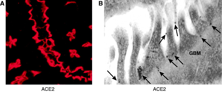
Figure 1.
Immunofluorescence and immunogold analysis of angiotensin-converting enzyme 2 (ACE2) in the kidney. (A) Immunofluorescence staining of ACE2 (red) in proximal tubules. (B) ACE2 immunogold labeling in glomeruli. ACE2 labeled with 15 nm of gold particles is distributed in podocyte foot processes and slit diaphragm (A, arrows). The glomerular basement membrane (GBM) does not have ACE2 immunogold particles (modified from ref. 19 with permission).

