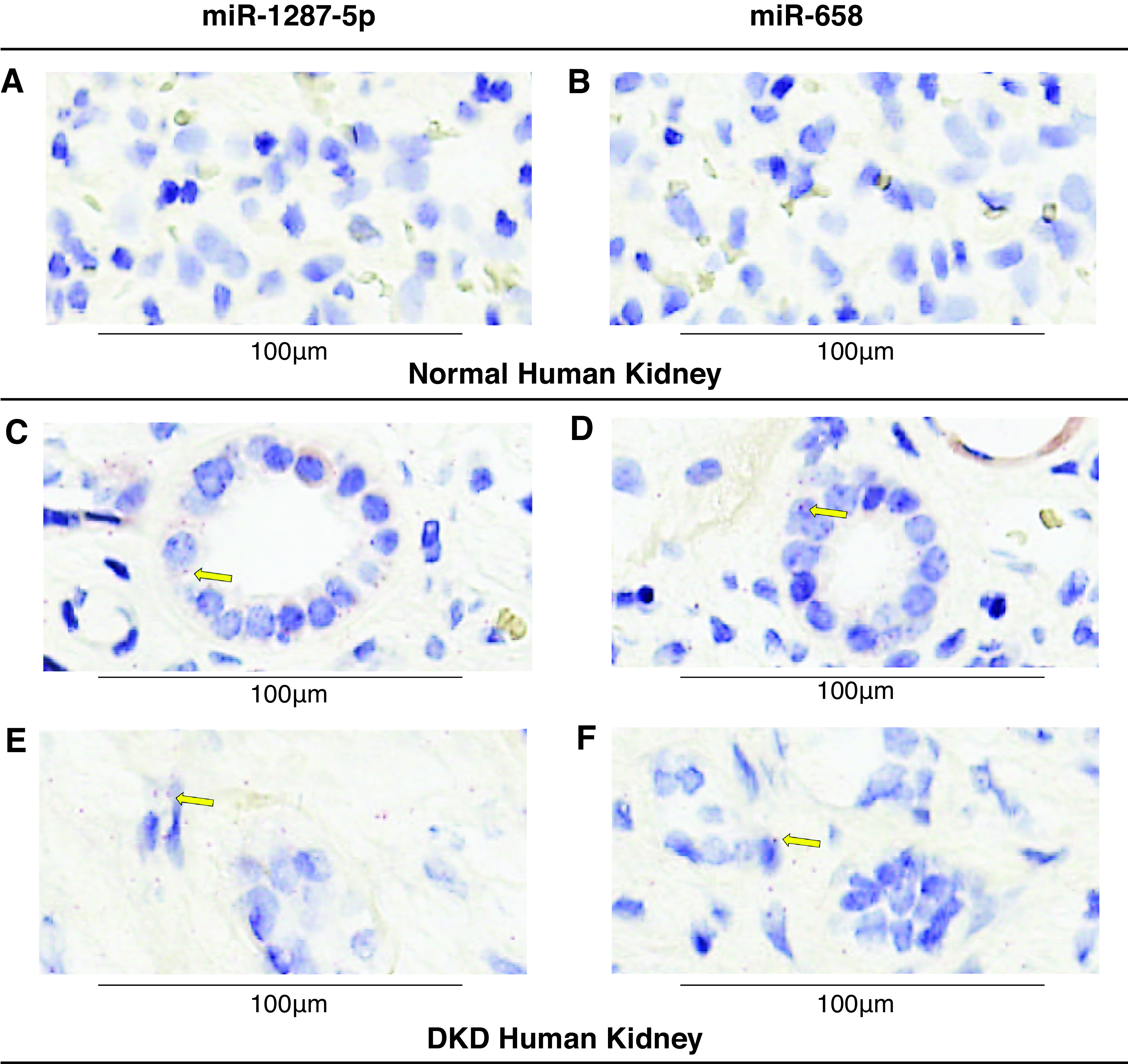
Figure 5.

Tissue expression of miR-1287-5p and miR-658 in normal and DKD human kidney samples using miRNAscope ISH assay. (A–B) miR-1287-5p and miR-658 ISH assays in normal human kidney. (C–F) Detection of miR-1287-5p and miR-658 in DKD kidney. Red punctate dots indicate positive staining marked by yellow arrows. Blue hematoxylin counterstains the nuclei. Original magnification, 40×. Scale bar, 100 μm.
