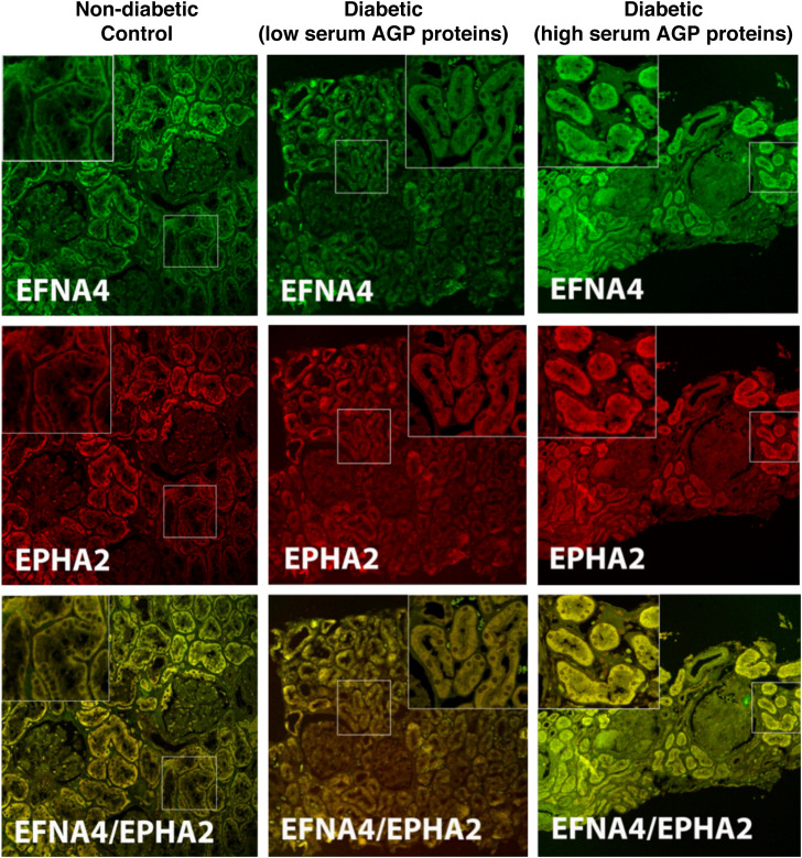
Figure 6.
Immunofluorescence staining for EFNA4, EPHA2, and colocalization of the two AGP proteins in kidney biopsy tissue. A nondiabetic, age-matched control (left column), a Pima subject with T2D (middle column) and low serum AGP proteins (EFNA4, 2150 relative fluorescent units [RFU]; EPHA2, 3453 RFU; and VvMes, 12.3%), and a Pima subject with T2D (right column) and high serum AGP proteins (EFNA4, 2333 RFU; EPHA2, 4462 RFU; and VvMes, 27.2%).

