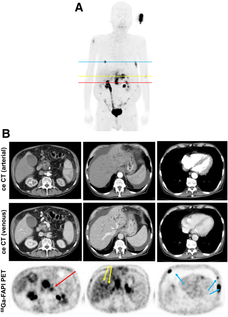
FIGURE 3.
Staging of patient with local recurrence of PDAC. (A) Mean intensity projection (MIP) image of FAPI PET/CT imaging. (B) Axial ceCT and FAPI PET/CT images of same patient on level of local recurrence (red line in A), 2 metastasis-suspicious intrahepatic foci (yellow line in A), and 3 suspicious osseous tracer accumulations (blue line in A). In contrast to CT imaging, FAPI PET/CT allows discrimination of metastatic lymph node from local recurrence mass (red arrow). FAPI PET/CT also revealed possible new liver (yellow arrows) and bone (blue arrows) metastases.

