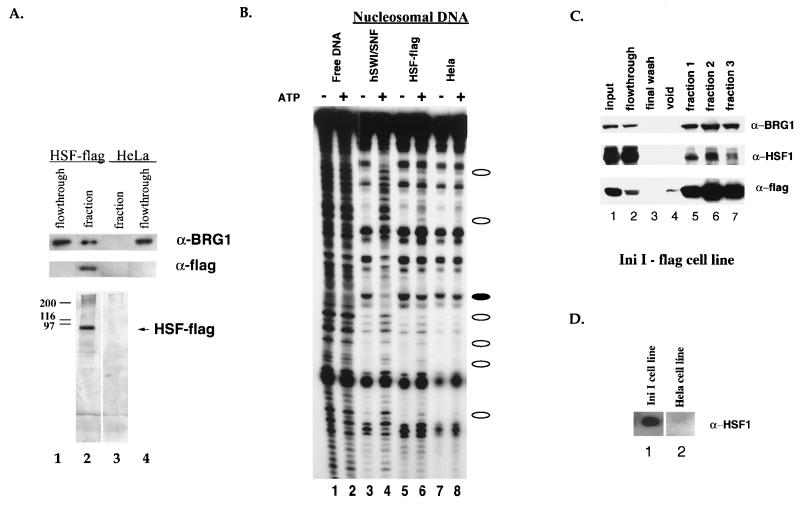
FIG. 2.
Association of human HSF1 with human BRG1 and chromatin remodeling activity. (A) Fractions from the M2 (anti-FLAG) purification of full-length HSF1-FLAG were resolved by SDS-PAGE and assayed by silver staining (bottom panel) or Western blot analysis using the anti-BRG1 antibody (top panel) and the anti-FLAG antibody, D-8 polyclonal (Santa Cruz) (middle panel). BRG1 flowthroughs are comparable to inputs. Heat shock fractions from cell lines expressing HSF1-FLAG (lane 2) and a HeLa cell line control (lane 3) were compared. (B) The same fractions from panel A were compared (lanes 5 to 8) to purified hSWI/SNF (lanes 3 and 4) in the DNase I mononucleosome disruption assay. Naked DNA was digested by DNase I as a control (lanes 1 and 2). Open ovals designate bands with an ATP-dependent increase in intensity, and closed ovals designate bands with an ATP-dependent decrease in intensity. Lanes 1 and 2 are at a lighter exposure in order to better see the bands in the highly digested lanes. (C) Fractions from an M2 (anti-FLAG) purification of INI1-flag, a hSWI/SNF subunit, were resolved by SDS-PAGE and assayed by Western blot analysis using the anti-BRG1 antibody (top panel), the anti-HSF1 antibody (Affinity BioReagents) (middle panel), and the anti-FLAG antibody (M5, Sigma) (bottom panel). The anti-HSF1 experiment required a longer exposure in order to visualize the HSF bands better. (D) Fractions from an M2 (anti-FLAG) purification of INI1-flag (lane 1) and a HeLa cell line control (lane 2) were resolved by SDS-PAGE and assayed by Western blot analysis using the anti-HSF1 anti-body (Stressgen).

